Lid watching, a lost art!


images-2

The diagnosis of blepharospasm is not always easy, in particular in case of pretarsal blepharospasm, also called Apraxia of eyelid opening, mistaken quite often for ocular myasthenia.

1- what is ocular myasthenia?

Myasthenia is an autoimmune disease, responsible of muscle weakness, affecting the all body and when localized to the muscles around the eyes, called ocular myasthenia.

Ocular myasthenia can present with isolated droopy eyelids (medically called ptosis), due to a weakness of the levator muscles of the eyelids, the Levator Palpebrae, but is often associated with double vision, due to weakness of the oculomotor muscles. The weakness is worst at the end of the day, due to the fatigue of the myasthenic muscles.

The diagnosis of ocular myasthenia is based on clinical findings, the presence of antibodies in the serum (30 to 60% cases) and the abnormal response of the muscles on repetitive electrical stimulation. Clinically the doctor can look for the “peek sign” by asking the patient to perform a sustained gentle eyelid closure; the fatigue of the orbicularis oculi will be responsible of a slight opening of the eye, as the patient peeking.! ( J. Glaser).

2- why so many patients with Blepharospasm are misdiagnosed with ocular myasthenia?

-Blepharospasm patients can have a misleading presentation of ptosis, with intermittent droopy eyelids, due to the spasms of the pretarsal portion of the orbicularis oculi muscles (also described as pretarsal BSP) pulling down the eyelid like the string of a roller blind down to the window.

-The words that patients used to describe their symptoms such as “my eyes are tired”, “my eyelids feel heavy”, “I feel more comfortable eyes closed” can indicate wrongly a muscle weakness of the eyelids, and reflect in fact the loss fight of the patients against the dystonic spasms closing their eyes.

– the variability of the symptoms through the day ; in myasthenia the patient is worst at the end of the day; in BSP the patient is worst walking outdoors, driving, watching TV, looking up and with any bright light and dazzy winter light.

– A levator palpebre detachement can occurs on some patients after years of spasms, and pulling on the muscle insertion

3-the art of lid watching

The famous neuro-ophtalmologist, J Glaser talked about “ Lid watching, a lost art!” Joel Glaser in Handbook of neuro-ophthalmology, 1999

-Lid watching is optimal when patients are keeping silent; BSP patients have an increased rate of blinking when being quiet and vice versa a low blinking rate when speaking. It’s the opposite of what observed in normal subjects. That may explain why patients find easier to keep her eyes opened when speaking and choose to sing when driving.

-In the case of pretarsal BSP careful lid watching along the eyelashes, looking for a pulling down spasms of the eyelids is a good indicator of pretarsal spasms.

– In addition, the lid closing spasms may be associated with Bell’s phenomenon with elevation of the eyeball, so it’s important to look not only at the eyelid but also the eyeball

– In case of levator palpebre detachment, the forceful opening of the eyes on command will be limited;

– Also the patients sometimes can’t reopen their eyes, after a spontaneous blink or after a closing spasm. It can be for a fraction of second or for up to few minutes; the patient will flicker his eyelashes, or his eyelids to kick them opened. Sometimes it will be a forceful pulling of the eyelids with his fingers, stretching the skin around the eyes, even sometimes resulting in bruising.

All these signs found on careful lid watching are good indicators of eyelids spasms, and not eyelid droopiness. They also direct the hands of the injector to the maximum spasms to achieve optimal results.

In the movement disorders field, most of the diagnosis are made by watching the movement; it’s particular true for eyelid dystonia and vocal cords dystonia where immobility does not always equal paralysis but also permanent tension due to the dystonia; one day I will ask my ENT colleagues to tell us about the art of vocal cords watching!!!
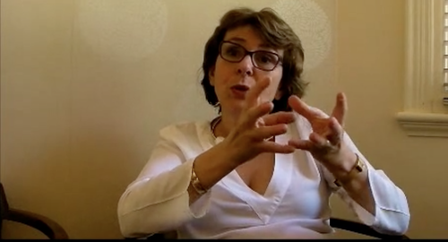
Marie-Helene Marion


Understanding dystonia and its treatment. Video on questions & answers

A good knowledge of the dystonia is important for a patient to become actor of its treatment. The Dystonia society UK asked me to answer to the most frequent questions coming from their members. Please follow this link if you want to watch this video posted on the Facebook pageof the Dystonia society.

image

The questions were as follow:

What causes dystonia in adults? Can any vaccine, peripheral nerve damage, the menopause or the surgery has any affects? (Time 1.00)

Does dystonia spread? If so, are there any particular parts of the body it spreads to and from? (Time 6.20)

How long do the symptoms carry on getting worse? (Time 7.58)

I understand Botulinum toxin is a type of poison. Is it safe to use? (Time 13.35)

Can Botulinum toxin be used in pregnancy? (Time 13.39)

Is there a way to avoid side effects from Botulinum toxin such as pain at the injection sites, swallowing difficulties, blurred vision? (Time 15.21)

How can I manage pain in dystonia if it is not relieved by Botulinum toxin? Is it OK to take pain medications on long term basis? (Time 18.57)

Why is my Botulinum toxin not working? Might is this because I become immune to the toxin? (Time 20.41)

Is Artane (Trihexyphenidyl) helpful? Is there an age limit for taking it? (Time 31.54)

Can physiotherapy or mind retraining help? (Time 36.50)

Might diet or complementary therapy help to manage dystonia? (Time 39.09)

I hope it will contribute to a better understanding of dystonia by the patients themselves and help them to cope better in their daily life.

Dr MH Marion

My jaw is out of control when speaking or chewing: what does it mean?

                                     image

                                                    the Iron jaw in circus

Eating is a simple pleasure of life and a necessity. Biting into a biscuit could require a tremendous effort when the movements of the jaw become out of control. The automatic movements of the jaw and the tongue, which allow us to eat or speak, can be disrupted by involuntary jaw spasms.


What are the causes of jaw spasms?

1-Idiopathic jaw dystonia is the most common cause of jaw spasms. The onset is between the age of 50 to 60 and it is more frequent in women. Dental works can trigger it. It’s called idiopathic dystonia as no underlying disease can be found. A genetic mutation has been identified in familial form of jaw dystonia ( DYT6).

2-Tardive jaw dystonia can follow a treatment with drugs used for the treatment of psychosis, called neuroleptics.

3- Hereditary disease affecting the brain is often the cause of jaw spasms occuring in young people, under the age of 20.

4- Hemi-masticatory spasms is usually a consequence of radiotherapy of the jaw area for cancer of the ENT sphere. In that case, the spasm is painful and affecting one side of the jaw with sudden, unexpected painful clenching of the jaw.

Are there different types of jaw dystonia?

1-The jaw spasms can be closing spasms with sudden clenching, responsible of tongue biting, teeth breaking and limitation to open the mouth wide

2-The jaw spasms can be opening spasms, responsible of difficulties to keep the mouth closed and to keep the food into the mouth. Often the tongue is involved and has a tendency to poke out the mouth.

3- The jaw spasms can also move involuntary the jaw side to side, or on one side only, or forward (protrusion) or backward (retrusion)

The movement involved in eating and speaking are incredibly complex and the dystonic spasms can be a combination of opening, deviation to one side and going backward or forward.

What are the characteristics of jaw dystonia?

The jaw spasm occurs in any attempts of eating and/or speaking, therefore the diagnosis requires looking at the patient performing these tasks. The doctors should have a box of biscuit available to examine their patients with dystonia!

The spasm are relieved by some tricks like keeping a sweet in the mouth or a chewing gum, sucking a matches or the temples of their spectacles.

How to treat jaw dystonia?


The most efficient treatment is the Botox injections of the masticatory muscles.

            -The muscles, which close the jaw, are the masseters, the temporalis and the median pterygoid muscles

image

Masseters, closing jaw muscles

image

Temporalis, closing jaw muscles

Figure adapted from Travell and Simons’  

-The muscles, which open the mouth, are the lateral pterygoid and the mouth floor muscles.

image

Mouth floor muscles ( opening jaw muscles), also called supra-hyoid muscles

Figure adapted from Travell and Simons’

image

Median ( closing jaw muscles) and lateral pterygoid (opening jaw muscles)

Figure adapted from Travell and Simons’

‘Some of these muscles are superficial and easy to inject ( masseters, temporalis,mouth floor muscles), some are deeper (median an lateral pterygoid muscles) and requires to be injected with electromyography guidance.

The difficult part is to analyse the abnormal movements and understand which muscles are involved in the dystonia. It can take many injections sessions, to get the spasms under control. The 2 limiting factors are the swallowing difficulties, due to the spread of the Botox , in particular when the tongue  muscles have to be injected.  Starting with small doses and increasing gradually the dosages is advisable.

The human masticatory muscles are very strong, in particular the muscles which closed the mouth; just think of the acrobats in a circus who get suspended by biting a mouthpiece. But at least injecting jaw muscles in human is possible if we compared them to the jaw clamping muscles of the crocodiles, which are extremely strong, and as hard as bone.

image

 Strong closing jaw muscles of the crocodiles

Dr Marie-Helene Marion is a specialist in Botox treatment for jaw spasms, and in particular for jaw and tongue dystonia.

British Neurotoxin Network 2013 meeting, Keble college, Oxford

image

 The 3rd British Neurotoxin Network annual meeting was hold in Oxford last week; 74 Botulinum toxin injectors gathered for all over UK in the superb location of Keble college. The program covered both practical and scientific aspects of botulinum toxin and dystonia.

 I gave a pre dinner talk on facial expression of emotions, looking on how the concepts have evolved from the 19th century with Charles Bells ‘s anatomical dissection of the facial muscles, to the 21st century with the influence of facial Botox injections on our expression of emotions.The collaboration of Duchenne de Boulogne with his neurophysiology study and Charles Darwin on expression of emotions in man and animals contributes to the present understanding of the universality of expressions and the recognition of basic emotions on a face, which was confirmed by Paul Ekman, an American psychologist.The Botox has been used as a research tool in the 21-st century to understand the relation between emotion and facial expression. Repressing the expression of emotions by too much cosmetic Botox or by neurological conditions such as facial palsy could disturb in return our ability to perceive emotions.

image      Duchenne de Boulogne, 1862

 imageCharles Darwin, 1872

The following morning was dedicated on reviewing the techniques and indications of the treatment of drooling by Ms Helen Witherow, maxillo-facial surgeon in London, the treatment of jaw dystonia by myself and the comparison of 2 techniques of injection: either electromyographic guided or ultra sound guided injection by Dr Sabine Klepsch , neurophysiologist in Bristol. Ms Catharina Pearce, medical student from Cardiff presented the results of a national survey on the use of Botulinum toxin in pregnant women.

In the afternoon the lectures were focusing on new concepts in dystonia and in particular the attempt of defining a new endophenotype (heritable clinical markers) for adult onset focal dystonia by Dr Sean O’riordan from Dublin and Dr Richard Grunewald from Sheffield.

 Outside the Botulinum toxin treatment, Mr Alex Green, neurosurgeon in Oxford, explored the effect of Deep Brain Surgery on dystonia and Mr Richmond Stace physiotherapist in London, the basis for retraining of cervical dystonia.

A video session, where colleagues were presenting video of dystonic patients with unexpected outcome, was animated and the opportunity again to share our individual approach to treatment.

The meeting was also a great opportunity for the secretary Mr Bells to update the UK maps of the service using botulinum toxin for treating neurological conditions. it’s accessible to anybody who is looking for a specialist centre to be treated for dystonia in particular.

 Dr Marie-Helene Marion, chair of the British Neurotoxin Network

Management of arm spasticity in Liverpool, 9th December 2011


Management of arm spasticity in Liverpool

9tH December 2011

 Last Friday,  I went to a Masterclass of  Management of arm spasticity organized by Professor Paul Mc Arthur hand plastic surgeon and Ms Jane McPhail, consultant maxillo-facial prosthetist.

It amazed me how much I always learn from colleagues from other specialties.. We all learn the same functional anatomy of a given muscle, so what’s make the   view so different  between a neurologist, treating a writer’s cramp and a hand surgeon treating a spastic arm?

 Spasticity and dystonia have a different origin. Spasticity in adults commonly followed a stroke with lesion of the cortex (the skin of the orange!). Writer’s cramp is a focal dystonia, which is due to a circuit problem between the deep nuclei (Basal ganglia-the seeds of the orange!) of the brain. Oppenheim, 100 years ago defined the word dystonia as an increased tone different from what was seen after stroke.

 The difficulties encountered by these patients are very different.

Dystonic patients such as Writer’s Cramp have problems when writing, but otherwise are fine for other tasks and at rest. Patient with spastic arms have a permanent disability at rest with pain, discomfort and abnormal posture, interfering with every action requesting skills and strength.

 The abnormal posture has nothing in common…the spastic arm present with rigid flexion of the elbow, arm in pronation (palm of the hand twisted and looking to the floor), and flexion of the wrist and fingers . In case of a dystonic arm, the abnormal posture is not fixed and will be present only when writing. Sometimes  the dystonia is more severe and the arm has a mobile, variable abnormal dystonic posture when arm outstretched and when walking; the whole arm is  twisted in pronation but without a fixed flexion of the elbow, the wrist in flexion and the fingers can be flexed or extended, animated with slow movements.

But despite all these differences, they both benefit from Botulinum toxin injections…

So the approach of the treatment has to be adjusted to the condition


Spastic arm: I learned from professor McArthur the concept of function unit to understand at best the spastic arm, with the biceps, brachialis and brachioradialis for the arm flexion, the pronator teres and the flexor carpi radialis for the arm flexion and pronation, and the synergy between the lumbricals and the flexor digitorum  communis . Also I had the demonstration that ultra sound can be crucial to target the spastic muscles precisely as the arm is permanently in an abnormal posture, modifying normal anatomical reference.  The electromyogram is not helpful, except the stimulation, as the voluntary command of the arm is poor.

 Dystonic arm: My experience of watching and treating dystonic patients taught me, on the other hand, that dystonic arm requires to be assessed during the task which triggered the dystonia, that 1 or 2 muscles in spasms can lead the all arm in a dystonic spasms, and that electromyogram is extremely helpful to document the dystonic bursts in the arm; the selection of the muscles is complex as the posturing is variable , but targeting the muscle itself under e;ectromypogram is not as difficult.

 Confronting experience and developing concepts between clinicians from different specialties, are invaluable, and the Liverpool course confirmed that looking from another perspective brings new ideas.

 Also spending a night in Liverpool at the Hard Days Night hotel, having breakfast surrounded by the Beatle’s portraits make you feel very far from London.

Marie-Helene Marion

London BTX centre

Professor Paul McArthur training course:

http://www.prostheticinnovations.com/surgicalworkshops.php

Sweating in Winter: a serious problem


Sweating in summer on a hot day or after a long Sunday jog may feel good, or at least normal. But sweating in winter when the weather is cold or you just come out of the shower can be a nightmare for individuals suffering from a condition called Hyperhydrosis (also spelled Hyperhidrosis).


Primary Hyperhydrosis or excessive sweating is in third of the cases a familial condition, which can have various manifestations, depending of the site of the excessive sweating; “primary” means that no obvious cause such as drugs, diabetes, infection etc.. is associated with the excessive sweating. Excessive sweating, in particular starting in adulthood and without family history should be investigated by the GP as it could be secondary sweating.

Armpits sweating:

 In case of Primary Hyperhydrosis affecting  the armpits, it’s often  young adults complaining of sweat marks on their clothes under their arms since their teens. They avoid colourful and white clothes and prefer dark clothes to not show the marks; they feel ashamed and loose their confidence as they are wrongly convinced that the excessive sweating under the arms is associated with excessive body odours. They bring extra clothes at school or at work to change during the day.

Hand sweating:

It can also affect the hands, usually noted when the child learned to write at the age of 5; the pen slipped out the hand; they made wet mark on the paper when writing. then these children don’t dare holding hands of their friends at primary school and later on avoid social contacts; it can also affect  professional manual skills.

Feet Sweating:

The excessive sweating of the feet is usually well tolerated in winter where the socks in the shoes absorb the sweat and I have met patients who were convinced that to have wet socks were normal at the end the day. The worst is in summer when ladies try to wear open sandals, in particular with some heels; the sweaty wet feet slip out and make walking hazardous. I have heard alos young men distress about having wet feet in bed.

Facial sweating:

Facial sweating is also very distressing. The sweat can drip from the scalp, or the forehead along the face, the cheeks and the nose and in back of the neck leaving the hair wet.

Excessive sweating people can benefit from treatment and is not related to excessive shyness or anxiety but it’s a true condition.

Please don’t hesitate to contact support group such as the Hyperhidrosis Support Group at www.hyperhidrosisuk.org

Dr MH Marion is running specialized clinics for all types of primary hyperhidrosis both at St George’s Hospital, London (UK) and at the  London BTX centre.

So many different types of focal dystonia!

So many different types of focal dystonia!

 Depending of the body part affected,

 ·      Eye closure is called Blepharospasm (blepharo means eyelid in Greek).

·      Jaw opening or clenching is called Oro-mandibular dystonia (oro means mouth and mandibular means jaw in Latin).

·      Tongue protrusion is called Tongue dystonia

·      Twisting of the head to one side is called Cervical dystonia (cervic means neck in Latin).

·      Writing difficulty is called Writer’s cramp.

·      Back arching, or trunk bending forward is called Axial dystonia (axial means axis as the spine).

·      Walking or running difficulty is called the “ Crampe du marcheur” in France!

·      Difficulty playing a musical instrument is called Musician’s cramp.

·      Speaking difficulty is called Spasmodic dysphonia (phonia means the speech).

 They usually start in adulthood and remain focal, without spreading to adjacent part of the body.

H.Oppenheim, 100 years ago was the first to use the word “ Dystonia”…

H.Oppenheim, 100 years ago was the first to use the word “ Dystonia”…what does the word Dystonia means? Dr Marion will take you back 100 years ago…

Dystonia is a neurological condition, characterized by involuntary sustained pulling of the muscles in one part of the body (focal dystonia, mainly in adult) or spread in the all body (generalized dystonia, mainly in children) and associated with abnormal postures.

The word “Dystonia” is composed of Dys (meaning abnormal) and Tonia ( meaning the tone ) . The tone represents how flexible or stiff is a part of the body. Oppenheim coined the word “Dystonia” in 1911 to describe an abnormal muscle tone, different from what was observed following stroke (spasticity). Oppenheim described children, from Ashkenazi Jewish descent, affected with generalized dystonia and called this condition “ Dystonia musculorum deformans”. In 1989, Laurie Ozelius established that a gene (called DYT1) on chromosome 9 was responsible of the ‘Oppenheim dystonia”.

 http://en.wikipedia.org/wiki/Hermann_Oppenheim

http://brain.oxfordjournals.org/content/97/1/793.extract

http://www.ncbi.nlm.nih.gov/pubmed/2576373

Cervical dystonia: what does the examination look for?

Cervical dystonia: what does the examination look for? Dr Marion tells you how the clinical examination is important to optimise the Botox treatment.

Cervical dystonia: what does the examination look for?

 Patients with cervical dystonia described at onset an intermittent feeling of pulling in the neck muscles; over the months, the head take an abnormal posture. Torticollis is defined by the head (the chin) turned to one side; laterocollis by the head tilt toward one shoulder; retrocollis by the head pulled backward in extension, chin up; antecollis by the  head  flexed downward, chin down to the chest. These abnormal postures are due to the involuntary movement of the neck on the trunk. There is also the possibility of abnormal movement of the head on the neck, the head going forward like a goose or going backward giving a double chin posture.

The diagnosis of cervical dystonia is made on the involuntary movement and the abnormal posture of the head, and often delays by many years from the onset of symptoms. They are other rare causes of abnormal posture of the head that a neurologist can exclude by a neurological examination and investigations.

 But like the BSP, every patient is different and need to be examined carefully to document precisely the abnormal dystonic posture to know which muscle is pulling too much! Then these muscles, which are responsible of the pulling, will be the one to inject with Botulinum toxin.  The difficulty comes from the action of the neck muscles which are often mixed (rotating the head on one side and tilting on the other side), resulting in mixed abnormal posture of the head (predominant rotation with a degree of tilt and extension).

It’s best to see the patient at his worst!

The patient is asked to walk, to stand up, to write or to lie down depending of the triggering factors. Usually the worst posture is achieved standing, eyes closed. The fixation of the eyes on an object helps the person to keep the head straight; when closing the eyes, the head lost the visual cue and shift to its maximal abnormal posture. Then to write down the degrees of the rotation, tilt, extension and flexion to be able at the next visit to assess the improvement under treatment.

It’s also important to see the patient at his best!

I ask the patient if he has a “geste antagoniste” (French expression also used by the Anglo-Saxons neurologists!); for instance stopping the pulling in rotation by touching with one finger the cheek without exerting an opposing force to the movement. What’s count is the improvement of the pulling by simply touching a part of the head or neck. It’s also called sensory trick and it’s a hallmark of the dystonic phenomenon.

Then when the posture is clearly documented, identifying which are the leading muscles behind, is based on surface anatomy (palpation) and functional anatomy (which muscle is doing what!).