Sensorimotor retraining for patients with dystonia with Richmond Stace and Sally Glover

Richmond Stace ( London) and Sally Glover (Birmingham), neuro-physiotherapists share their large experience of treating patients with dystonia in this webinar organised by Dystonia UK. Dystonia is often a life changing condition which require for the patient to move forward and reflect about his own goals and expectations. Relearning how to move and live with the dystonia is a journey that Richmond and Sally will take you through in this enlightening webinar.

Webinar with Richmond Stace and Sally Glover on Dystonia

Retraining of Cervical Dystonia with Jean Pierre Bleton, organised by Dystonia Europe

Webinar Dr JP Bleton and dr MH Marion on 9th May 2020, organised by Dystonia Europe, with Adam Kalinowski and Edwidge Ponseel

Thank you to Adam Kalinowski, president and Edwidge Ponseel, vice president of Dystonia Europe for organising this webinar about the retraining of patients with Cervical dystonia.

Thank you to Jean Pierre Bleton for providing this invaluable educating tool for patients understanding better what ‘s happening in their neck muscles and what to do to control better the dystonic spasm. It will be also very helpful for physiotherapists to access the unique experience of JP Bleton on this topic.

Dystonia in pandemic

Patients with dystonia have difficulties accessing Botulinum toxin clinics at this time of pandemic.

I have recorded a video presentation to advise dystonic patients with recommendations if the injections are not available.

Dr Marion Dystonia in Pandemic – 30/03/20

No panic ! a lot can be done.

I hope you will find this message helpful

All my best wishes Dr Marie-helene Marion

Other webinars written by colleagues on “what to do in time of Pandemic”

1-Voice Techniques to Manage Symptoms of Spasmodic Dysphonia and Related Voice Conditions

2-On exercises to manage dystonia without Botulinum toxin

by my Italian colleagues, Anna Castagna , Elisa Andrenelli e Marina Ramella

An international blogger network


DMRFC_BloggerBadges_fin-10

The Dystonia Medical Research Foundation (DMRF) Canada is a Canadian registered charity and their mission is to advance research for more treatments and ultimately a cure for dystonia; to promote awareness and education; and to support the needs and well-being of affected individuals and families.

As part of their mission, they provide an international blogger network. I am delighted to be part of this network and to share information about dystonia and its treatment with Botulinum Toxin on this blog.

A better understanding  of the dystonia helps the patient to become actor of its treatment  and to develop the confidence that the patient needs to successfully carry out a specific behaviour in a given situation (self efficacy), a key to minimise disability due to the dystonia.

Clear written information about treatment and possible side effects (swallowing difficulties, double vision and droopy eyelid) is part of a well informed consent procedure to treatment and allows the patient to face the side effects when they occur, with appropriate management and less anxiety.

Networks are not beneficial for patients but also for doctors! I have founded in 2011 the British Neurotoxin Network to share information and best practice between UK injectors for neurological indications. I also look forward meeting again my Canadian colleagues from Quebec for a dystonia day in May 2018.

Lid watching, a lost art!


images-2

The diagnosis of blepharospasm is not always easy, in particular in case of pretarsal blepharospasm, also called Apraxia of eyelid opening, mistaken quite often for ocular myasthenia.

1- what is ocular myasthenia?

Myasthenia is an autoimmune disease, responsible of muscle weakness, affecting the all body and when localized to the muscles around the eyes, called ocular myasthenia.

Ocular myasthenia can present with isolated droopy eyelids (medically called ptosis), due to a weakness of the levator muscles of the eyelids, the Levator Palpebrae, but is often associated with double vision, due to weakness of the oculomotor muscles. The weakness is worst at the end of the day, due to the fatigue of the myasthenic muscles.

The diagnosis of ocular myasthenia is based on clinical findings, the presence of antibodies in the serum (30 to 60% cases) and the abnormal response of the muscles on repetitive electrical stimulation. Clinically the doctor can look for the “peek sign” by asking the patient to perform a sustained gentle eyelid closure; the fatigue of the orbicularis oculi will be responsible of a slight opening of the eye, as the patient peeking.! ( J. Glaser).

2- why so many patients with Blepharospasm are misdiagnosed with ocular myasthenia?

-Blepharospasm patients can have a misleading presentation of ptosis, with intermittent droopy eyelids, due to the spasms of the pretarsal portion of the orbicularis oculi muscles (also described as pretarsal BSP) pulling down the eyelid like the string of a roller blind down to the window.

-The words that patients used to describe their symptoms such as “my eyes are tired”, “my eyelids feel heavy”, “I feel more comfortable eyes closed” can indicate wrongly a muscle weakness of the eyelids, and reflect in fact the loss fight of the patients against the dystonic spasms closing their eyes.

– the variability of the symptoms through the day ; in myasthenia the patient is worst at the end of the day; in BSP the patient is worst walking outdoors, driving, watching TV, looking up and with any bright light and dazzy winter light.

– A levator palpebre detachement can occurs on some patients after years of spasms, and pulling on the muscle insertion

3-the art of lid watching

The famous neuro-ophtalmologist, J Glaser talked about “ Lid watching, a lost art!” Joel Glaser in Handbook of neuro-ophthalmology, 1999

-Lid watching is optimal when patients are keeping silent; BSP patients have an increased rate of blinking when being quiet and vice versa a low blinking rate when speaking. It’s the opposite of what observed in normal subjects. That may explain why patients find easier to keep her eyes opened when speaking and choose to sing when driving.

-In the case of pretarsal BSP careful lid watching along the eyelashes, looking for a pulling down spasms of the eyelids is a good indicator of pretarsal spasms.

– In addition, the lid closing spasms may be associated with Bell’s phenomenon with elevation of the eyeball, so it’s important to look not only at the eyelid but also the eyeball

– In case of levator palpebre detachment, the forceful opening of the eyes on command will be limited;

– Also the patients sometimes can’t reopen their eyes, after a spontaneous blink or after a closing spasm. It can be for a fraction of second or for up to few minutes; the patient will flicker his eyelashes, or his eyelids to kick them opened. Sometimes it will be a forceful pulling of the eyelids with his fingers, stretching the skin around the eyes, even sometimes resulting in bruising.

All these signs found on careful lid watching are good indicators of eyelids spasms, and not eyelid droopiness. They also direct the hands of the injector to the maximum spasms to achieve optimal results.

In the movement disorders field, most of the diagnosis are made by watching the movement; it’s particular true for eyelid dystonia and vocal cords dystonia where immobility does not always equal paralysis but also permanent tension due to the dystonia; one day I will ask my ENT colleagues to tell us about the art of vocal cords watching!!!

Marie-Helene Marion


Mental imagery and dystonia

Mental imagery is already used in high level sports competition, and athletes are trained to rehearse their race in their mind, to increase their performance.

Could dystonic patients benefit from mental imagery focusing on their motor
difficulties
?

There are scientific facts, and anecdotal reports from dystonic patients to suggest that it may be very helpful on the long term.

Real and mentally stimulated movements rely upon largely overlapping networks. In another words, writing or imagining writing involved common networks in our brain. Studies have shown that the mental imagery of a movement in patients with dystonia follows the same difficulties than the execution of the dystonic movement.

A study by Fioro, 2006 has looked at the mental rotation of body part (hands, feet) in which the subjects imagine moving their body part, from their actual posture into that seen on a picture to recognise which side is belonged in patients with writer’s cramp. Writer’s cramp patients are slower than controls in mentally rotating hands but not feet; it suggests that the mental rotation of body parts reflects the anatomical constraints of real hand movements.

Could the mind reshape the brain?

Neuroplasticity is a normal process by which the brain develops new connections at different levels, following interaction with environment, emotions, behavior. Dystonia is understood to be a condition characterized by an excessive neuroplasticity of the brain leading to abnormal learning of motor program. To be able to influence this brain plasticity It’s important to create an environment for positive learning and recovery.  Mindfulness can be helpful in patients with focal dystonia to minimise stress and increase well being before embarking for retraining.

Retrain your brain, not your muscles!

The visual and mental imagery training will encourage you to imagine successful, normal execution of tasks, which the dystonia prevents you to do..

Remember the time when you could perform the movement normally, and how easy it was to do it and the pleasure, which come with it.

It will be best performed after the Botox treatment has already controlled the dystonic spasm
in the affected body part and together with sensory retraining.

Visualisation exercises are not always easy and you may benefit of the advice of professionals such as hypnotherapist or specialized physiotherapist

Try daily to visualize yourself free of the dystonia, walking eye opened, turning your head freely looking around you, writing a page with our favorite pen or chewing a delicious meal.

Also music and melody can help you to IMAGINE…

You may say I’m a dreamer, But I’m not the only one, I hope someday
you’ll join us, And the world will live as one  (John Lennon)

Speaking up about silent swallowing difficulties in dystonia

Ms Lucy Hicklin ENT, Mr Paul King chief executive Dystonia society UK, Dr Marie-Helene Marion neurologist and Carol Harris speech therapist. London living with dystonia event, on swallowing and speech problems on the 8th november 2014, London.

Every day we swallow solid food like a piece of bread, water and our own saliva. This vital function occurs most of the time without us being aware of it, as a reflex. But in fact swallowing is a very fine and precise mechanism, which can be disrupted in a number of neurological conditions, the most common and dramatic one occurring after a stroke, but also in dystonia.

A few definitions:

1-    Doctors talks about swallowing difficulties as dysphagia, which is the difficulty moving food from mouth to stomach.

2-    Swallowing difficulties can be severe leading to a misdirection of the food to the nose, or to the larynx (voice box) and the lungs, resulting in infection of the lungs called aspiration pneumonia (as the food has been sucked int the airways).

3-    Swallowing difficulties are silent when there is no manifestation which will make the person aware of the swallowing problem, such as cough, choking, needed to drink to push down the food.

4-    Swallowing difficulties are subclinical when the signs are so mild that they are overlooked such as cutting food into small pieces and taking a sip of fluid with each swallow.

 What are the essentials for good swallowing?

1-    Of course the machinery such as  the tongue, the mouth, the throat (the pharynx) the voice box (the larynx), the oesophagus (gullet) have to be healthy. Issues apart from neurological problems, such as a tumour, could obstruct the swallowing process.

2-    The nervous system has to be able to conduct in an orderly fashion the different sequences of the swallowing, like a conductor with his musicians; only one “ fausse note” can disturb the all process. It’s called neurogenic dysphagia.

What are the sequences of the swallowing?

image

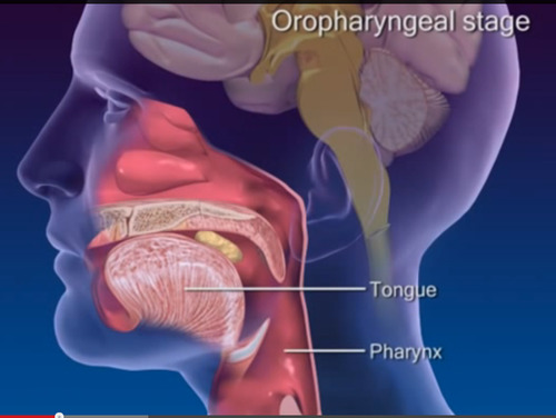
1-    The oral phase: the food is collected in the mouth as a soft mass of chewed food, called a bolus.

2-    The pharyngeal phase: the swallowing reflex is triggered by the bolus touching the back of the throat, which give a signal to the brain ( cortex and brainstem) to start the safe guard process: the airway needs to be protected by elevating and closing the larynx and by lifting the soft palate to close the back of the nose. The door to the oesophagus (the crico-pharyngeal sphincter) opens to let in the bolus which has been propelled downwards by the back of the tongue and the pharyngeal muscles.

3-    The oesophageal phase. The bolus then passes though the gullet and into the stomach. This is under the control of the autonomic nervous system

How can swallowing difficulties be assessed?

1-    -During a consultation, a speech therapist will examine the tongue strength, gag reflex, voluntary cough, and speech and perform a trial of swallowing (water, puree) and/or a 3 ounces (90 ml) water test.

2-     FESS (fiberoptic endoscopic evaluation of swallowing).

A fine “spaghetti “( fibre-optic) tube, with a camera at the end, connected to a screen, is introduced by the nose and passed gently down to the throat. With the camera in place the patient is asked to swallow a food of different consistencies.   The food is normally dyed blue or green to make it easy to see against the pink of the gullet.  The speech therapist performing the test can see where it is going, particularly if it is getting stuck or going down into the airway (aspiration) .

3-    Videofluoroscopy is video-radiography of the mouth, throat and oesophagus when the patient swallows a range of foods (biscuit, puree, yoghurt) and liquids that are coated in barium.  The barium shows up on the x-rays (radio-opaque). On the video the radiographer can follow the trajectory of what has been swallowed.

4-  Recording the swallowing process with electrodes in the muscles under the chin and movement sensors on the Adam’s apple can give a good indication of the timing of the different swallowing phases.  It’s called an electrophysiological assessment.

image

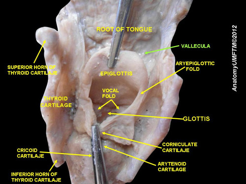
Posterior aspect of the base of tongue and larynx , after removing the pharynx http://commons.wikimedia.org/wiki/File:Slide10vvv.JPG#mediaviewer/File:Slide10vvv.JPG

What about swallowing difficulties in dystonia, in untreated patients?

Very few studies have addressed this topic. Most have concentrated on the swallowing difficulties in dystonic patients following treatement with  Botulinum toxin injections.

I will present 5 studies of swallowing difficulties in patients with cervical and craniocervical dystonia, before treatment.

1-Riski et al (1990) studied 43 patients with cervical dystonia with videofluoroscopy and found that 51% of the patients had swallowing difficulties; in 16% the difficulties were subclinical (not known by the patient). The patients had mainly a delay in triggering the swallowing reflex and some residue in the vallecula (a groove, behind the base of tongue, usually cleared by the posterior movement of the base of tongue).

2Comella et al (1992) studied 18 patients with cervical dystonia with videofluoroscopy before and after Botox injections; 11% patients had clinical symptoms of swallowing difficulties and 22% had radiologic signs of peristaltic abnormality.. after Botox (see below), the signs and symptoms of dysphagia in these patients did not change, but an additional 33% developed new dysphagic symptoms and 50% of the patients developed new peristaltic abnormalities by radiologic studies.

3- Munchau et al (2000) studied 12 patients with cervical dystonia undergoing selective peripheral denervation with videofluoroscopy. 90% patients had swallowing abnormalities with 58% subclinical. The patients presented as in the Riski’s study, with delayed initiation of the swallowing reflex and abnormal tongue base posterior movement, in particular in patients with saggital shift of the head.

The subclinical dysphagia was mainly cutting food into small pieces and taking a sip of fluid with each swallow.

4Ertekin et al (2002) studied 25 patients with cranio-cervical dystonia with electrophysiological assessment. . 36% had clinical dysphagia and 72 % had abnormal swallowing on electrophysiological recording with again delayed swallowing reflex ; also the sphincter ( the crico-pharyngeal muscle) which opens the oesophagus was hyperactive, instead of being relaxed during swallowing to let the food going through.

Ertekin developed the concept of the dysphagia limit. In this simple test patients are asked to swallow volumes of water ranging from 1ml to 20mls.  In patients with clinical swallowing problems only 1-10 mls can be swallowed in one go. In subclinical patients 10-15mls can be managed.  All normal subjects could swallow 20mls in 1 gulp.

5-Cersosimo et al (2005) studied 20 pts with Meige syndrome with videofluoroscopy. 90% patients had abnormal swallowing compared to 40% in a healthy population of same age. They found also residue in the vallecula as in Riski’s study

What about swallowing difficulties in dystonia in Botox dystonic patients?

Botulinum toxin injections, by weakening adjacent muscles, have been responsible of transient dysphagia in dystonic patients.

The different types of dysphagia depending of the sites of the Botox injections have been detailed in a previous post.

Conclusion

Swallowing difficulties are often silent or subclinical in dystonic patients. Patients and doctors need to speak about them as these swallowing difficulties can be worsened by Botulinum toxin treatment administered for the treatment of the dystonia.

Maybe every injector needs to offer a drink to their dystonic patients before treating them! It may be well received…..

image


 References: 

Swallowing function in patients with spasmodic torticollis.; Riski JE1, Horner JNashold BS JrNeurology. 1990 Sep;40(9):1443-5

Dysphagia after botulinum toxin injections for spasmodic torticollis: clinical and radiologic findings.; Comella CL1, Tanner CMDeFoor-Hill LSmith C.; Neurology. 1992 Jul;42(7):1307-10

Prospective study of swallowing function in patients with cervical dystonia undergoing selective peripheral denervation; A MunchauC GoodS McGowanN QuinnJ Palmer, and K Bhatia; J Neurol Neurosurg Psychiatry. Jul 2001; 71(1): 67–72.

Oropharyngeal swallowing in craniocervical dystonia, C Ertekin*I Aydogdu**Y SeçilN KiyliogluS TarlaciT Ozdemirkiran J Neurol Neurosurg Psychiatry 2002;73:406–411

Swallowing disorders in patients with blepharospasm.: Cersósimo MG1, Juri SSuárez de Chandler SClerici RMicheli FE. Medicina (B Aires). 2005;65(2):117-20

http://www.infodystonia.com/post/24333066534/swallowing-difficulties-after-botox-injections-what-to


Understanding dystonia and its treatment. Video on questions & answers

A good knowledge of the dystonia is important for a patient to become actor of its treatment. The Dystonia society UK asked me to answer to the most frequent questions coming from their members. Please follow this link if you want to watch this video posted on the Facebook pageof the Dystonia society.

image

The questions were as follow:

What causes dystonia in adults? Can any vaccine, peripheral nerve damage, the menopause or the surgery has any affects? (Time 1.00)

Does dystonia spread? If so, are there any particular parts of the body it spreads to and from? (Time 6.20)

How long do the symptoms carry on getting worse? (Time 7.58)

I understand Botulinum toxin is a type of poison. Is it safe to use? (Time 13.35)

Can Botulinum toxin be used in pregnancy? (Time 13.39)

Is there a way to avoid side effects from Botulinum toxin such as pain at the injection sites, swallowing difficulties, blurred vision? (Time 15.21)

How can I manage pain in dystonia if it is not relieved by Botulinum toxin? Is it OK to take pain medications on long term basis? (Time 18.57)

Why is my Botulinum toxin not working? Might is this because I become immune to the toxin? (Time 20.41)

Is Artane (Trihexyphenidyl) helpful? Is there an age limit for taking it? (Time 31.54)

Can physiotherapy or mind retraining help? (Time 36.50)

Might diet or complementary therapy help to manage dystonia? (Time 39.09)

I hope it will contribute to a better understanding of dystonia by the patients themselves and help them to cope better in their daily life.

Dr MH Marion

Why Spasmodic dysphonia is often mistaken for a functional disorder?

                                                 image

Spasmodic dysphonia, also called laryngeal dystonia, is a neurological disorder, responsible of a strangulated, strained voice and rarely of a whispering voice, which has a major negative influence at work and in personal life.

Patients with spasmodic dysphonia require to be distinguishing from patients with functional dysphonia and from patients with “muscle tension dysphonia” (MTD) which is also considered a functional disorder rather than a neurological disorder.

Many reasons can explain why spasmodic dysphonia , which is a dystonia of the larynx, is not well recognized by doctors.

1- Laryngeal dystonia is a rare condition (1/100 000), and like other focal dystonia, the diagnosis is clinical; if you have never heard somebody with spasmodic dysphonia speaking, it will be very difficult to recognize it.

2- It mainly occurs in women, who carry the stigma of being more vulnerable for functional disorders.

3- The onset can be sudden in 45% of the cases (Childs, 2011).  Sudden onset is also a characteristic of functional disorders.

4- When the onset is sudden, the majority of patients recognized triggering factors such as stress (42%), upper respiratory infection (33%), and pregnancy and parturition (10%).(Childs, 2011). Stress should be seen as a non-specific triggering factor making patients vulnerable to develop any medical conditions, and not specifically functional disorders.

5-A phobic component occurs very quickly after onset, as the patient avoid answering the phone, and speaking in public as it’s required such an effort to speak in these circumstances.

6-It’s a task specific condition as every focal dystonia,; spasmodic dysphonia occurs when speaking, and voice can be normal for singing or shouting. This task specific characteristic can be disconcerting for both the patient and the doctor.

At the difference, functional dysphonia is responsible of a permanent speech disturbance, with a whispering voice or a hoarse voice occurring when speaking, and impossibility of shouting or singing.

7- “Muscle tension dysphonia” (MDT) patients tend to exert too much effort on their vocal cords when speaking and can present with a strained voice, as spasmodic dysphonia.

But when examining the speech in details, three tasks: sustained vowel /a/, oral reading of a standard passage, and connected speech describing a standard picture have to be performed, as the spasmodic dysphonia could be more apparent in the connected speech situation, compared to MDT.

                                          image

It’s very important to diagnose spasmodic dysphonia, as Botox injections into the vocal cords can alleviate the symptoms and restore confidence for effortless speaking.

At the London BTX centre, in Sloane Square, Dr. Marie-Helene Marion offers multidisciplinary clinics with voice specialists for the diagnosis and  treatment of spasmodic dysphonia with Botox injections.